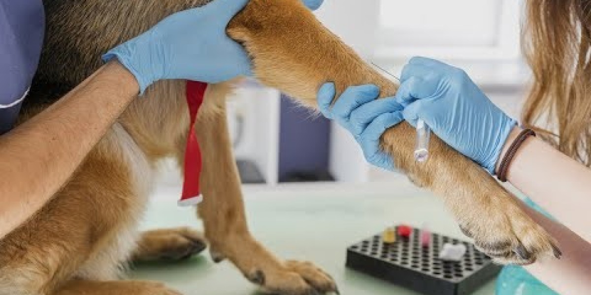
How to receive reproductive health services at the DNC
Here's what you should find out about heart disease in canines, tips on how to recognize the indicators, and ways to help prevent coronary heart disease so that your canine can stay his greatest, happiest life for a few years to return.
 Right ventricular enlargement results in rounding and cranial enlargement of the cranial cardiac border on the lateral film and rounding of the best cardiac border which is nearer to the thoracic wall than normal on the VD film . In some cases this produces the reverse D appearance on VD or DV radiographs. A systematic approach to evaluating the mediastinum is crucial to establishing normality for a given small animal. One can consider roentgen abnormalities of the mediastinum as both major (an abnormality of the mediastinum itself) or secondary (an abnormality caused by a mediastinal construction or organ). Examples of primary mediastinal abnormalities would include abnormal fluid or fuel collections inside the mediastinum. Examples of secondary mediastinal abnormalities would include lymphomegaly, cardiac abnormalities, esophageal problems, tracheal issues and irregular hemorrhage of tumors of the thymus. The first two net primarily based reviews contain identifying normal airways of the canine thorax on a right lateral and on a ventrodorsal radiograph.
Best Dog Toothbrushes: Traditional, Dual-End, Three-Sided, Electric & More (& Our Personal Experience)
Because the act of respiration changes the thoracic look, inspiratory movies ought to be attempted to distinguish artifacts from true lung pathology. High-quality, 3-view thoracic movies which are appropriately positioned ought to be the beginning point for all practitioners. For greatest results, the practitioner must be actively concerned in processing the photographs to ensure right anatomical positioning and publicity (Figure 1). Whenever attainable, analises clinicas veterinaria more than 1 person ought to evaluation films for abnormalities and roentgen changes. Interpretation of thoracic radiographs may be difficult because most disease processes often involve several areas of the thorax.
Radiographic Anatomy
If a structure appears "abnormal," the abnormality must be categorized based on the roentgen signal method because greater than 1 change may be current, depending on the disease process. When reviewing films, the clinician should suppose in phrases of radiographic opacity and use the standard roentgen signal strategy. Radiographic opacities permit the interpreter to distinguish steel (white), mineral, delicate tissue, fat, and air (black). Expiratory (A) and inspiratory (B) right lateral radiographs from a 7-year-old neutered male poodle.
This reference doesn't suggest any intention on the part of the Editor to sell such product, application or service in the User's nation. The Site and the Applications include data on services that may or will not be out there in all countries of the world. The User must guarantee, previous to any use of the Site, analises Clinicas veterinaria the Applications or the products/services accessible through the Site and the Applications, that such use does not violate the legal guidelines of his nation of residence. In basic, we advocate that you simply systematically search the recommendation of your ordinary physician before consulting any Internet websites and functions with medical content material. The Customer could access the contracts binding the events at the time of the order (Subscription Conditions, Privacy Policy, Terms and conditions of access and use of the Site and Applications) on the Site and by way of links throughout the Applications. The Customer may download these contracts and hold them in a durable manner on some other medium of his alternative.
What Does a Chest X-ray Reveal in Dogs?
On every lateral radiograph, a dorsal fissure is situated round T6 between the cranial and caudal lung lobes. On lateral views, the ventrally oriented lobe bronchi could also be troublesome to visualise (especially the proper middle bronchus on a proper lateral radiograph). The proper cranial lung lobe bronchus exits the carina dorsal to the center base and turns 90° in a ventral and cranial course (FIGURE 4). The left cranial lung lobe bronchus exits the ventral trachea at the carina (FIGURE 4). Within roughly a centimeter from its origin, the left cranial lobar bronchus divides into the cranial and caudal segmental bronchi. Just caudal and ventral to the carina, the best middle lung lobar bronchus originates from the ventral and lateral side of the proper caudal lobe bronchus.