La insuficiencia cardiaca en el perro se define como la incapacidad del aparato circulatorio como consecuencia de una cardiopatía para conseguir que la sangre se distribuya por todo el organismo y garantizar su preciso desempeño.
Los dóbermans, por otro lado, son especialmente susceptibles a la miocardiopatía dilatada, una afección que desgasta las paredes del corazón y disminuye de manera significativa su capacidad de bombear sangre de forma eficaz.
Pulse deficits are sometimes because of a untimely beat that occurs so early that the ventricles are unable to fill sufficiently, resulting in a decreased stroke volume that produces both a weak pulse or no pulse.
 Determine the Anatomic Source of the Rhythm
 The diaphragm may contract synchronously with the guts to provide loud thumping noises on auscultation and often seen contraction in the flank area. The syndrome outcomes from stimulation of the phrenic nerve by atrial depolarization and happens primarily when there is a marked electrolyte or acid-base imbalance, particularly with hypocalcemia. Synchronous diaphragmatic flutter is most common in horses and canines. In dogs it happens mostly in association with hypocalcemia and electrolyte disturbances induced by GI disease, though idiopathic circumstances also occur.
Heart rhythm
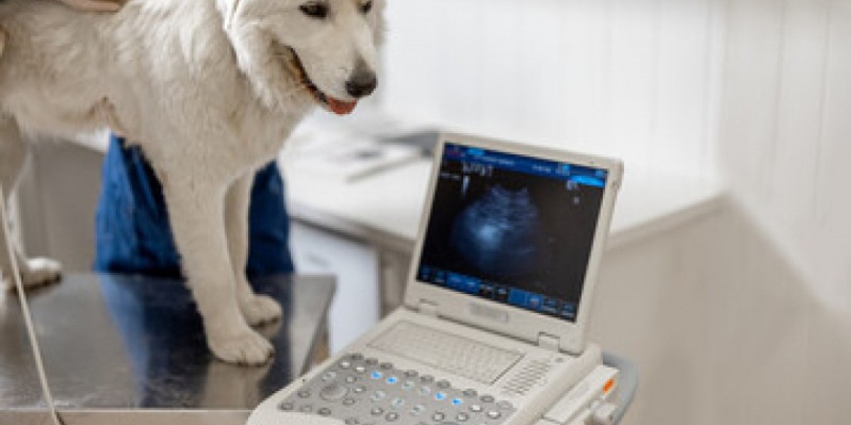
Advanced imaging, similar to magnetic resonance imaging, Laboratório clínico veterinário is now being used more usually to diagnose the presence of cardiac lots in sufferers with pericardial effusion. If your vet has determined your pet wants more thorough cardiac monitoring, your pet might be referred to a cardiologist, who will probably have him put on a Holter ECG. The Holter is an ambulatory monitor that wraps around your pet’s torso comfortably and stays on for twenty-four to seventy two hours. The take a look at results are recorded and sent to the cardiologist for analysis. Your pet can proceed his usual routine with out disruption while carrying the Holter.
Why does my pet need an echocardiogram?
He is a charter member of the Academy of Internal Medicine for Veterinary Technicians and served on its government board representing cardiology for 12 years. Previously, laboratóRio Clínico Veterinário he spent 18 years at the University of Missouri within the cardiology service and 3.5 years at Ross University School of Veterinary Medicine within the anesthesia service. He has a ardour for instructing, is the editor of Cardiology for Veterinary Technicians and Nurses, and has written over a dozen peer-reviewed articles. It is your responsibility to confirm that any CE course accomplished through the Sites qualifies for CE credit in your state.
A Practical Approach to Sustainability in the Veterinary Clinic
A single lead would supply info on just one dimension of current move. For the needs of this overview, we’ll give attention to lead II, a bipolar lead during which the right arm (RA—the right foreleg in veterinary patients) is adverse and the left leg (LL—or left hind leg), is constructive (Figure 1). Before starting any interpretation, first observe what is termed CLAP (the amplitude Calibration, Lead displayed, any Artifact, and Paper speed). This step shortly identifies the presence of bradycardia or tachycardia. Because HR is a component of cardiac output, an irregular HR can have a deleterious effect on cardiac output. Decreased cardiac output may be noted as hypotension in the affected person. Monitors could display HR for the operator, but these values should be considered with scrutiny as a end result of the HR algorithm might incorrectly calculate HR because of artifact, arrhythmias, or excessively giant ECG waveforms.
The first step in diagnosing heart disease in dogs is a complete physical examination. The veterinarian will use a stethoscope to take heed to your dog’s heart and lungs to check for irregular rhythms and feels like coronary heart murmurs or crackles (evidence of fluid in the lungs). X-rays (also referred to as radiographs) of the chest incessantly help diagnose coronary heart illness in pets. Finding generalized enlargement of the guts or enlargement of particular coronary heart chambers makes the presence of coronary heart disease more likely.


